

The shoulder
The anatomy of the provides the strength and functionality of the upper body. It is a complex structure of bones, muscles and ligaments with the ability to lift weights and create enormous strength. The shoulder can counteract an extreme impact but is also vulnerable to to a range of pathologies due to inactivity, overuse and trauma. Repetitive action also also sees an increase in wear and tear of the connective tissue causing pain, limited mobility and structures degradation.
The shoulder is one of the most complex joints of the body as it allows for almost a 360 degree motion. In its complex function, the shoulder must be mobile enough to allow for the wide range of actions of the arms and hands, but also stable enough to execute lifting, pushing and pulling. The compromise between mobility and stability may result in a large number of shoulder conditions that are less frequent in other joints of the body.

Bones of the shoulder
The shoulder consists of the head of the humerus fitting in to the glenoid fossa, a depression of the scapula (shoulder blade). These two parts fit with each other to form a socket-ball joint. These bones are supported in the anterior part of the body by the clavicle, or collarbone, and in the posterior side by the acromion, the roof of the scapula. The cartilage covering the shoulder joint confers structural support to reduce friction of the articulating elements and facilitate a wide range of movements.

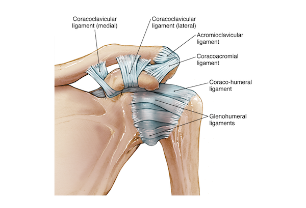
Shoulder joints and ligaments
The movement of the shoulder is permitted by three distinct joints:
acromioclavicular (AC) joint formed by the acromion, which is the postero-anterior bone of the scapula and the clavicle bone
sternoclavicular (SC) joint situated at the centre of the upper chest right under the throat where both clavicle bones meet (breast bone)
glenohumeral joint is the main joint of the shoulder consisting of the humeral head inserting in the glenoid.

The shoulder capsule
To keep the head of the humerus within the glenoid, a robust yet flexible connective tissue forms the shoulder capsule, which is the major ligament system of the shoulder. The capsule is formed by the glenohumeral ligaments and encloses the entire shoulder joint. It attaches the upper part of the humerus to the shoulder blade.

The glenoid labrum is a fibro-cartilaginous elastic structure that encloses the glenoid cavity creating a deep socket where the glenohumeral joint rests. At the superior side note the attachment to the long head of the biceps brachii muscle. The glenoid labrum is essential to provide stability to the shoulder joint.
Rotator cuff
The ligaments’ pivotal role is to connect bones to other bones, whereas tendons connect muscles to bones. The rotator cuff is an assembly of tendons originating from the muscles subscapularis, on the anterior side and the supraspinatus, infraspinatus and teres minor (on the posterior side) joined together to form a cuff around the shoulder.

Together the muscles and tendons of the shoulder stabilise the upper arm bone, the humerus within the joint and are essential for the wide range of movement of the shoulder. Due to its particular structure and function, the rotator cuff is often subjected to injury and overuse. On the front side of the shoulder note the large subscapularis muscle with its tendon attached to the humeral head.

Subacromial bursa
The bursa is a fluid-filled sac enclosed in a synovial membrane. In the body there are several bursas that are intrinsic parts of the joints. The bursa is generally located within joints or beneath tendons to prevent friction of the tendons and bones during motion.
In the shoulder the subacromial bursa is situated between the acromion and the insertion of the supraspinatus tendon – one of the three tendons forming the rotator cuff – positioned above the capsule. It extends underneath the deltoid muscle and above the head of the humerus.
The subacromial bursa is one of the largest bursas of the body and is often subjected to various conditions most notably the impingement of the shoulder caused by the inflammation of the bursa, a pathology also named bursitis.

Muscles
The muscles of the shoulder provide stability to the complex elements forming the joint. Each muscle originates in the scapula and inserts to the humeral head to form a strong bond. The tendons of the muscles support the humerus during the movement of the shoulder such as rotation, adduction and abduction. On the superior aspect of the shoulder is the deltoid muscle and on the anterior side the coracobrachialis, serratus anterior, pectoralis major, and pectoralis minor muscles. These muscles work together to flex and adduct the scapulas and move the humerus anteriorly. The latissimus dorsi and teres major on the upper back are used to extend and adduct the arm towards the spine.

The trapezius, romboid major, and the levator scapulae muscles allow to raise the scapula when shrugging the shoulders and move the scapula towards the back.

The supraspinatus muscle is situated on the upper region of the shoulder just above the scapula and extends to the greater tubercle of the humerus bone. It is an important muscle as it is one of the four muscles forming the rotator cuff together with the infraspinatus, teres minor and subscapularis. The supraspinatus is used in shoulder abduction. The infraspinatus and the supraspinatus are separated by the spine of the scapula on the posterior side of the shoulder.The pectoralis minor is located below the pectoralis major muscle above the third, fourth and fifth rips.

Also on the posterior side of the shoulder is a pair of rhomboid muscles on either side of the spine. They are used to retract the shoulder. The teres major muscle is located above the latissimus dorsi muscle and is used when extending and rotating the humerus.

Arteries
The arteries supplying the shoulder arise from the heart, the aorta, and the subclavian artery. This artery is a large branch of the aorta exiting the heart to supply the upper arm. As it descends, the subclavian artery branches into the axillary artery that turns around the humeral head and the deep brachial artery that runs along the deeper structure of the arm.

Veins
The main veins of the shoulder, upper arm, elbow and forearm are the cephalic vein, the basilic vein, and the median cubital vein situated on the superficial upper extremities.

Nerves
The nerves of the arm originate from the cervical spine vertebrae (C5 to C8 and T1) and descend along the body to form the brachial plexus from where they interchange and extent along the shoulder, the upper arm and forearm up to the hand and fingers. The main nerves of the shoulder / upper extremities are the axillary nerve, the median, the radial and ulnar nerve.
The axillary nerve, also named the circumflex nerve, is located in the axilla and originates from the brachial plexus. It innervates the upper arm and shoulder region. Differently from other nerves it does not descend along the arm. After leaving the plexus it divides into the anterior and posterior branch. The anterior branch innervates the deltoid muscle in the upper arm while the posterior branch extends to the upper shoulder, supplying the teres minor (a muscle located in the lateral side of the scapula attaching to the rotator cuff in the shoulder joint) and the posterior area of the deltoid. The axillary nerve provides sensory function to the skin of the shoulder and deltoid muscle.

The median, radial and ulnar nerves
The other nerves of the upper extremities innervate the arm, wrist and hand. The median nerve controls the following motor functions: forearm pronation, thumb palmar abduction and thumb/index/long finger flexion. The motor branches of the radial nerve contribute to the function of the elbow, wrist, finger and thumb. The ulnar nerve controls fine hand movements, coordination of finger motion and pinch strength, flexion of the small and ring fingers.